


New Eye Cleaning System Discovered
The discovery of a previously
unknown drainage pathway
opens science’s eyes to more design
A New Eye Cleaning System Discovered
The human eye is more complex than once believed
by Jerry Bergman, PhD
Eyesight is essential to my work as an author, so I take my eye health seriously. I make a point of visiting my eye doctor regularly and following her instructions. One recent concern has been that my eye pressure was rising. If the pressure becomes too high, it increases the risk of glaucoma and damage to the optic nerve, potentially leading to permanent vision loss. When fluid does not drain efficiently, pressure inside the eye will often rise.
Pressure is one of the key factors associated with glaucoma, which often involves impaired drainage of aqueous humor and elevated pressure within the eye. However, glaucoma, age-related macular degeneration, and other causes of vision loss do not necessarily involve the same underlying processes or the same accumulation of fluid, waste, and inflammatory material. But eye pressure is only part of the story. Scientists have also been investigating how the eye removes waste and excess fluid to keep its delicate tissues healthy.
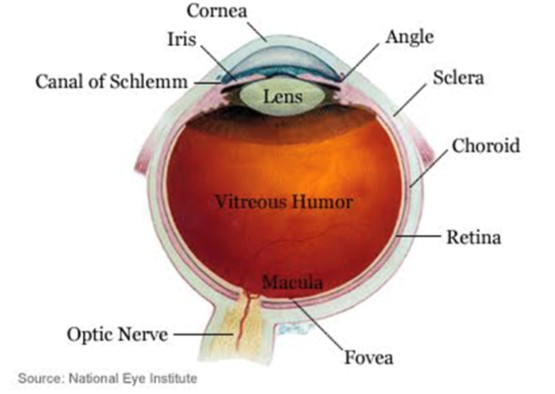
Figure 1. Illustration showing the location of the Schlemm’s canal. Image courtesy of the National Eye Institute (NEI).
How the Eye Normally Drains Fluid
The eye has a complex drainage system that helps regulate its internal pressure. The clear fluid called aqueous humor circulates through the front of the eye, supplying nutrients and carrying away waste products. Most of this fluid drains through a spongy tissue called the trabecular meshwork, located near the junction of the cornea and iris. From there, the fluid enters Schlemm’s canal (shown on the left in the accompanying eye diagram), then passes through small collector channels into veins that return it to the bloodstream. .
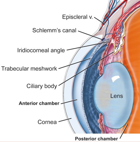
Fig 2 The location of the Ciliary body and Schlemm’s canal on the human eye. From Wikimedia Commons.
Aqueous humor is a clear fluid produced by the ciliary body of the eye. (see figure 2) The fluid helps maintain the required intraocular pressure and supplies nutrients to parts of the eye that lack their own blood supply. Maintaining a proper balance between the production and drainage of aqueous humor is essential for regulating eye pressure. When this balance is disrupted, intraocular pressure may rise, increasing the risk of glaucoma. Age-related macular degeneration (AMD) is related to eye health, thus eye pressure. This is another major cause of vision loss that affects approximately 40 million Americans and 2.5 million Canadians.[1]
The Research Methodology
To investigate how fluid drains from the back of the eye, researchers combined several advanced imaging methods including magnetic resonance imaging (MRI), near-infrared fluorescence imaging, and microscopic examination. They placed fluorescent tracer molecules into a narrow space at the back of the eye. By tracking the movement of these molecules, the researchers were able to follow the fluid’s pathway and determine where it drained.
How Eye Pressure Damages the Eye
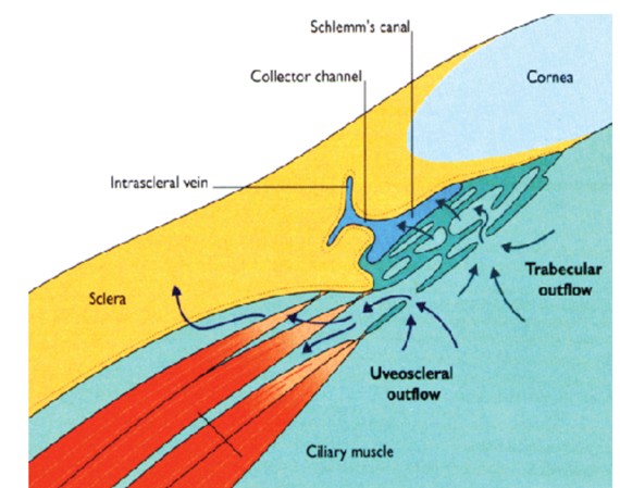
Figure 3 illustrates the uveoscleral pathway. Through this route, aqueous humor passes through the ciliary muscle and surrounding tissues before draining out of the eye.
High eye pressure (specifically intraocular pressure) damages the human eye primarily by compressing delicate nerve fibers and cutting off blood flow to the retina and other cells at the back of the eye. The high pressure inside the eye damages the optic nerve, the nerve which carries visual information from the retina to the brain. It is a major risk factor for glaucoma, which can damage the optic nerve and cause permanent vision loss if not adequately managed. To deal with this condition, my eye doctor prescribed Xalatan to help lower it. Xalatan (latanoprost) lowers eye pressure primarily by increasing aqueous humor outflow through the uveoscleral pathway rather than the conventional drainage route through the trabecular meshwork and Schlemm’s canal.
The New Discovery
Scientists have recently identified a previously unrecognized drainage pathway at the back of the eye.[2] Called the posterior ocular lymphatic outflow (POLO) pathway. To remove excess fluid and waste it carries fluid from the space behind the retina into the body’s lymphatic system. The discovery challenges long-standing assumptions about how the eye drains fluid and could eventually open new avenues for research and treatment of diseases that threaten vision. The research was conducted by Babishaa Sauntharrajan et al, at the University of Toronto Department of Laboratory Medicine and Pathobiology.[3]
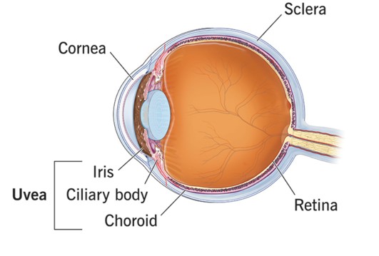
Figure 4 The Uvea Pathway of Aqueous humor drainage. Drawing from Cleveland Clinic
Excess aqueous humor leaves the eye through two well established drainage routes. The conventional pathway carries fluid through the trabecular meshwork and Schlemm’s canal into collector channels and, ultimately, to the episcleral veins. In contrast, the uveoscleral pathway allows fluid pass through the ciliary muscle and into the surrounding tissues before draining into the tissues of the eye’s outer layers and the eye orbit.
The Sauntharrajan et al team have now identified a third, previously unrecognized drainage route at the back of the eye. This system is similar to having two kidneys: if one fails or cannot handle the load, the other kidney can assist or take over. All three of these drainage systems function like backups to ensure that the required drainage occurs, given that the three routes function as interchangeable backups or work together to maintain fluid balance. Researchers have struggled to explain how this highly sensitive region of the eye clears excess fluid, metabolic waste, inflammatory material and other problematic substances.
The previously unknown drainage route was named the posterior ocular lymphatic outflow (POLO) pathway. This pathway carries both fluid and waste away from the back of the eye and into the body’s lymphatic network. The discovery reveals a previously unrecognized mechanism that may help regulate fluid balance and clear substances from this sensitive region. It could also provide new directions for investigating the eye’s immune defenses and diseases that threaten vision.
The retina is the light-sensitive tissue at the back of the eye. It requires substantial energy to convert incoming light into signals that the brain interprets, generating metabolic byproducts in the process. These byproducts must be removed to maintain healthy retinal tissue.[4] Dr. Neeru Gupta, professor and head of University of British Columbia’s department of ophthalmology and visual sciences, emphasized the significance of the discovery:
“The retina is one of the most metabolically active parts of the body, constantly generating byproducts that need to be cleared. This discovery helps to explain how the eye flushes this waste and promises to transform how we think about, and treat, a range of eye conditions.”[5]
By learning more about how the newly identified drainage pathway works—and what happens when it malfunctions—researchers will eventually discover new ways to protect vision and develop treatments for certain eye diseases.
This Discovery Challenges More Than a Century of Wrong Assumptions
For more than a century, scientists believed that the human eye lacked the type of lymphatic drainage systems found in most other body organs. (see figure 5). Throughout the body, lymphatic vessels collect excess fluid and help transport waste products and coordinate immune responses, helping maintain healthy tissues.
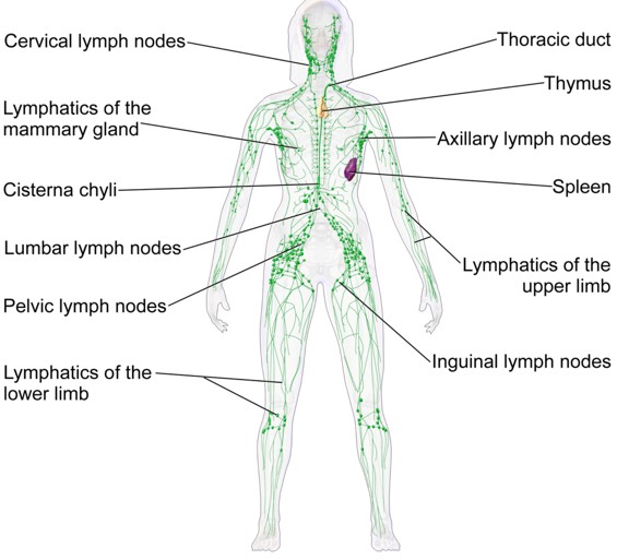
Figure 5 The body’s lymphatic system, which runs alongside the blood circulatory system. Image source: Wikimedia Commons.
Conclusions
The back of the eye has remained relatively poorly understood, even though the retina and surrounding tissues are affected by diseases that can cause permanent vision loss. However, the discovery of a previously unknown drainage pathway in this region will help researchers better understand how the eye clears excess fluid and waste—and what happens when this process is disrupted.
Scientists long believed that the eye lacked a conventional lymphatic vessel network within its tissues, the eye’s drainage mechanisms, particularly at the back of the eye. Each new anatomical discovery reveals another layer of complexity in the human body. This growing complexity raises further questions about how such intricate biological systems could have arisen through evolutionary processes involving genetic variation which is then acted upon by natural selection.
 Announcement: Dr Bergman’s new book, Whale Evolution Is a Whale of a Tale, is now available on Amazon. Subtitled “How a Dog Evolved Into a Whale” is a must-read for anyone interested in the topic of whale evolution. It exposes the claims of evolutionists Dr. Phil Gingerich and Dr. Hans Thewissen concerning supposed intermediate forms Pakicetus, Rhodhocetus and Ambulocetus. After these intermediates were shown to have been concocted, the evolution community has continued to crank out new theoretical land ancestors that they believe evolved into whales. Each new claim counters the previous claim. A feature that makes this book extra-special is all the fascinating background information on whale anatomy and physiology.
References
[1] The American Macular Degeneration Foundation. https://www.macular.org/about-us.
[2] University of British Columbia. “The eye has a hidden cleanup system scientists never knew existed.” ScienceDaily, 11 October 2026. <www.sciencedaily.com/releases/2026/10/261008005254.htm>.
[3] Babishaa Sauntharrajan, et al. 2026. Identification and Mapping of a Posterior Ocular Lymphatic Outflow (POLO) Pathway Through Choroidal Lymphatics. Translational Vision Science, 2026; 15 (7): 23 DOI: 10.1167/tvst.15.7.23.
[4] University of British Columbia. 2026.
[5] University of British Columbia. 2026.
 Dr. Jerry Bergman has taught biology, genetics, chemistry, biochemistry, anthropology, geology, and microbiology for over 40 years at several colleges and universities including Bowling Green State University, Medical College of Ohio where he was a research associate in experimental pathology, and The University of Toledo. He is a graduate of the Medical College of Ohio, Wayne State University in Detroit, the University of Toledo, and Bowling Green State University. He has over 1,900 publications in 14 languages and 40 books and monographs. His books and textbooks that include chapters that he authored are in over 1,800 college libraries in 27 countries. So far over 80,000 copies of the 60 books and monographs that he has authored or co-authored are in print. For more articles by Dr Bergman, see his Author Profile.